3D Precession Diffraction Tomography in TEM / case study
A 100 years old problem solved : the structure of vaterite
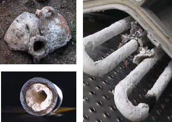
Vaterite, one of the common natural CaCO3 polymorphs, plays a pivotal role in weathering
and biomineralization processes. Vaterite is important for the problem of scales
in pipes, and for understanding biomineralization processes in mollusks and pearls.
Differently from calcite and aragonite (the other CaCO3 polymorphs), vaterite can
be found only in the form of nanosized crystals, not suitable for structure determination
by X-Ray diffraction. The structure of vaterite has eluded structure determination
for almost 100 years and is still an unsolved dilemma. We report here for the first
time an ab-initio determination of vaterite structure based on precession electron
diffraction (PED) data collected with using 3D diffraction tomography in a 300 kV
TEM.
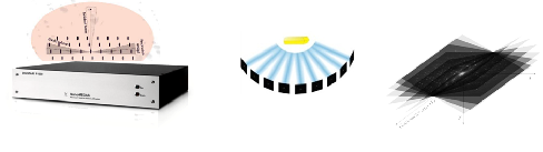
DigiSTAR – Precession electron diffraction device enables the collection of quasi-kinematical
intensities (X-Ray like) in any TEM. PED, in combination with the software ADT-3D
for 3D diffraction tomography, enables the reconstruction of the reciprocal cell
of any nanomaterial and the automatic integration of reflection intensities. Complete
3D diffraction data were collected from a single vaterite nanocrystal (50 nm or less).The
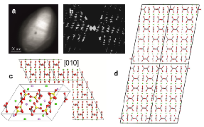
structure of vaterite was determined in monoclinic space group C2/c and is characterized
by a layer arrangement of Ca2+ ions alternated by {CO3}2- groups (2-layer model).
The closer analysis of vaterite nanocrystals showed stacking disorder and local modulation
which has been approximated by a 6-layer superstructure triclinic cell, resulting
in a perfect fit with synchrotron powder diffraction data. The superstructure was
solved ab-initio by direct methods in the triclinic space group C-1 .Electron diffraction
tomography showed its great potential to solve structure of nanomaterials that elude
conventional methods because of small crystal size and modulations.
Fig.1: (left) DigiSTAR precession device, (center and right) 3D diffraction tomography schematics with PED patterns collected around a common tilt axis.
Fig. 2 (a) STEM image of the crystal used for the acquisition of ADT/PED data set (b) 3D reciprocal space reconstructed from ADT acquisition, view down the tilt axis (c) [010] projection of vaterite 2-layer monoclinic model and (d) [010] 6-layer triclinic model (Ca atoms in green, C in grey, O in red).
Electron microscope: FEI Tecnai F30 –DigiSTAR (1.2°precession angle) - ADT-3D software, Fischione tomography holder (tilt -60/+60° in 1° step). Research Group: Institut für Physicalische Chemie der Johannes Gutenberg –Universität, Mainz, Germany. Ref: E. Mugnaoili et al., Angew. Chem. Int. Ed. 51, 7041-45 (2012).


 Vaterite, one of the common natural CaCO3 polymorphs, plays a pivotal role in weathering
and biomineralization processes. Vaterite is important for the problem of scales
in pipes, and for understanding biomineralization processes in mollusks and pearls.
Differently from calcite and aragonite (the other CaCO3 polymorphs), vaterite can
be found only in the form of nanosized crystals, not suitable for structure determination
by X-
Vaterite, one of the common natural CaCO3 polymorphs, plays a pivotal role in weathering
and biomineralization processes. Vaterite is important for the problem of scales
in pipes, and for understanding biomineralization processes in mollusks and pearls.
Differently from calcite and aragonite (the other CaCO3 polymorphs), vaterite can
be found only in the form of nanosized crystals, not suitable for structure determination
by X-